What Is a Lung Nodule?
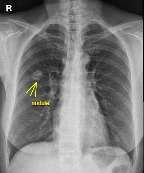
A lung nodule is a round, or oval growth in the lung, usually detected during routine imaging tests such as a chest X-ray or a CT scan. Most are less than 3 cm in diameter and larger ones are called masses. The discovery of a lung nodule can be quite alarming but not all of them are cancerous. Some studies estimate that up to 50% of all patients who undergo CT Scan of the lungs do have a nodule. In recent years, faster and better CT Scan technology have resulted in detection of more lung nodules than 20-30 years ago. Once a lung nodule is detected, patients usually need to seek the help of a respiratory physician for further evaluation. Some nodules are a sign of something serious, like infections, inflammation, or even early-stage lung cancer. Chest Xrays may not be able to detect small lung nodules and even with CT Scan, AI (artificial intelligence) is increasing being deployed nowadays to detect very small nodules.
Common Causes of Lung Nodules
These nodules can develop owing to many factors, such as:
- Infectious Granulomas: Granulomas are clusters of immune cells that are the most common type of benign nodules. Fungal lung infections or tuberculosis can cause granulomas to form.
- Non-Infectious Granulomas: Autoimmune diseases like rheumatoid arthritis and sarcoidosis can lead to the formation of non-infectious granulomas.
- Benign Tumours: These typically include hamartomas, lipomas, and adenomas.
- Cancerous Tumours: A common cause of pulmonary nodules includes non-small cell lung cancer, small cell lung cancer, and carcinoid tumours.
- Fibrotic Scarring: Smoking or inhaling lung irritants or chemicals can lead to lung scarring, which causes nodules.
What Indicates the Presence of a Lung Nodule
Most nodules do not cause symptoms, making early detection difficult. However, in cases where symptoms appear, they may include:
- Persistent coughing that lasts a few weeks
- Shortness of breath or difficulty breathing
- Wheezing
- Chest pain that becomes worse with deep breathing or coughing
- Coughing up blood (haemoptysis)
- Hoarseness
- Frequent respiratory infections such as bronchitis or pneumonia
- Unexplained weight loss
- Fatigue
When Should You See a Specialist?
Although most lung nodules are generally harmless, they should be generally be evaluated by a respiratory specialist who will take into account many factors,
- History of smoking
- Age and Sex
- History of prolonged exposure to harmful chemicals eg asbestos
- Family history of lung cancer
- The size of nodule
- Nodule growth rate over time
- Location of the nodule
- Nodule characteristics eg calcium, spiculated margin
- Number of nodules
- Any symptoms eg pain, loss of weight
Diagnosis and Testing
When a lung nodule is detected, the specialist may order a series of tests, which include:
- CT Scan with contrast: This imaging test provides detailed images of the nodule to assess its size, composition, and shape.
- PET Scan: This test helps determine whether the nodule is active, which may indicate potential malignancy.
- Bronchoscopy: A thin, flexible tube with a camera is inserted into the airways to collect tissue samples for an analysis.
- Needle Biopsy: A needle is used to extract tissue from the nodule.
Possible Treatment Options
The treatment approach for lung nodules may differ, depending on the patient and nodule’s characteristics and calculated risk of cancer. These include
- Surveillance: Active surveillance with follow up imaging to see if it grows or changes with time.
- Surgical removal: This is usually done when the risk of early lung cancer is significant
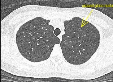
Case 1: 37 year old female non-smoker with father recently diagnosed lung cancer. Undergoes surgery for 7 mm ground glass nodule which turns out Stage zero lung cancer (early stage)
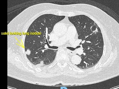
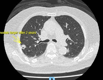
Case 2: 84 year old female non-smoker, comes for second opinion for lung nodule on CT seen 2 years ago. Follow up CT shows the solid looking nodule has grown from 7mm to 10 mm. Surgery was done and confirmed Stage 1 lung cancer.
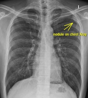
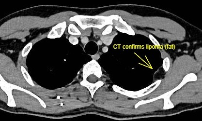
Case 3: 40 year old Male smoker asymptomatic was referred for an abnormal Chest Xray. CT confirmed pleural lipoma (fat on the skin of the lungs), a benign condition that does not require any further treatment or follow up.
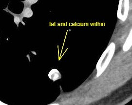
Case 4: 60 year old referred for lung nodule incidentally found on CT Scan. Repeat CT was done and images of the nodule magnified and windowed on the computer, confirming a hamartoma (benign) not requiring further treatment or followup.

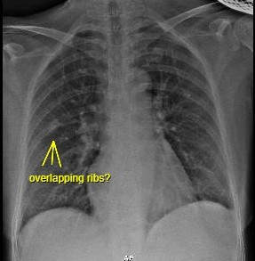
Case 5: 60 year old female being investigated for a right lung nodule seen on chest Xray. Review of previous Xrays a year ago already showed a possible nodule, thought to be due to overlapping ribs. Patient was advised to come back then for repeat CXR but defaulted. She was subsequently found to have stage 3 lung cancer.