|
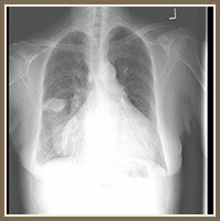
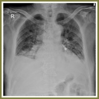
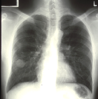
Chest X-ray showing “mass” in right lung due to fluid which resolved without thoracocentesis |
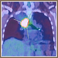
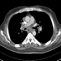
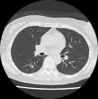
CT scan of thorax showing a tumour in the left pulmonary artery (simulating a blood clot/ pulmonary embolism) |
|
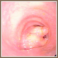
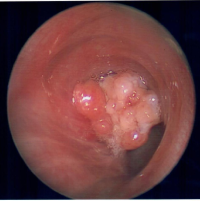
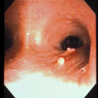
Bronchoscopic appearance of a rare benign tumour (lipoma) removed by bronchoscopy with Argon Plasma Coagulation |
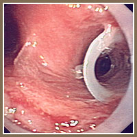
Stenting of the trachea and right main stem bronchus |
|
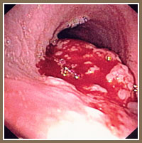
Rare lung cancer involving trachea; Biopsy showed mucoepidermoid cancer |
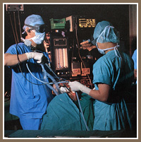
Flexible bronchoscopy under general anaesthesia |
|
Rigid bronchoscopy under general anaesthesia |
PET scan appearance of an unusual right lung tumour – turned out to be pulmonary lymphoma (a rare tumour in the lung) |
|
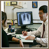
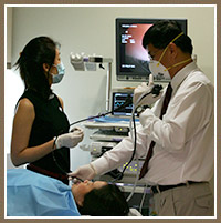
Consultation with Dr. Philip Eng |
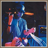
Flexible bronchoscopy under local anaesthesia |
|
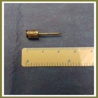
A foreign body (dental implant) lodged in the left lung |
Foreign body (dental implant) removal from lung via bronchoscopy |
|
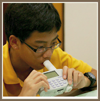
A 13-year-old undergoing a lung function test (Spirometry) and Methacholine Challenge test |
Coughing of blood due to aspergilloma (fungus ball) |
|
Cancer of oesophagus post-chemotherapy and radiotherapy (RT). Develops persistent cough and the above CT shows tracheoesophageal fistula necessitating bronchoscopy and stenting |
Rare lung tumor due to oncocytoma was removed via bronchoscopy (previously thought to be adenoid cystic cancer) |
|
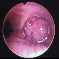
Uncommon airway tumour – Carcinoid tumour |
Carcinoid tumour – removed by bronchoscopy and laser |
|
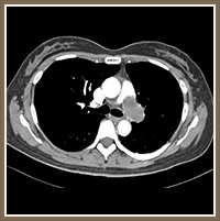
Right lung nodule which turned out to be stage 0 lung cancer |
|